ADH regulates water content of blood
ADH = Anti Diuretic Hormone
Found in a part of the brain (Hypothalamus)
Flows through the body until it reaches the kidneys
Has the ability to alter quantity of water in the blood
Therefore it can make the blood more or less concentrated
Describe the role of ADH in regulating the water content of the blood.
Targets the collecting duct and makes it more porous
.
Nature
วันจันทร์ที่ 7 พฤศจิกายน พ.ศ. 2554

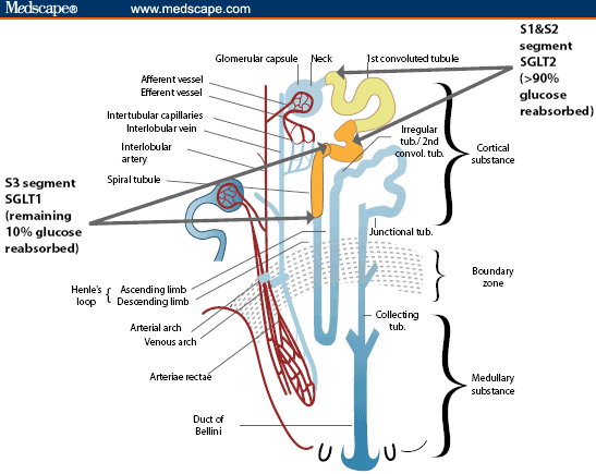
2.73 Glucose reabsorbtion
Similarly to the water some of the glucose is also lost
This means that it has to be re absorbed
this is done by the promximal convoluted tubule
If there is glucose in the urine however this means the person has diabetes

This means that it has to be re absorbed
this is done by the promximal convoluted tubule
If there is glucose in the urine however this means the person has diabetes
2.72 Water re-absorbtion
Ultra filtration sometimes filters out to much water
To prevent water loss the water must be absorbed back
This happens when the water passes along the collecting duct
The water is removed from the filtrate and is absorbed back into the blood vessels

2.71

Nephron carries out filtration of blood
Ultra filtration = filtration of molecules
Blood comes in and out of the arteriole and causes high pressure
This forces the plasma out of the blood vessel
The blood is filtered by utilizing high pressure
<iframe width="480" height="360" src="http://www.youtube.com/embed/VtCAUly4cdk" frameborder="0" allowfullscreen></iframe>
2.70 Structure of the Nephron

The lighter outer area is called the cortex
The dark shade in the middle is called the medulla
The light spot in the center is called the pelvic and is wear urine is stored
The bowman’s capsule is the dead end of a tubing structure in the kidney
The tubing structures are made up of twisted sections
A tight knot of blood vessels is known as the glumerous
Their are millions of Nephrons in a single kidney
They give the kidney its different colour tones
วันอังคารที่ 1 พฤศจิกายน พ.ศ. 2554
2.69

· The urinary system includes the kidneys, bladder, urethras and ureters.
· Each component has its own individual blood supply
· The ureters links the two kidneys to the bladder and transfers the urine
· The bladder is then linked to a structure called the urethra which carries the waste out of the body.
2.68b
· The kidney carries out a process called osmoregulation
· Osmo: osmosis & regulation: control
· Cells in the body are ideally ISOTONIC with the tissues around them
· This means that the amount of water passing in and out of the cells are equal and therefore the cells stay the same size
· However the blood that circulates in and out of the cell maybe of a different concentration and therefore cause a HYPERTONIC or a HYPOTONIC solution.
· Both of these solutions could harm the cells and therefore the most desirable option is an ISOTONIC solution.
· This is why the composition of the blood has to be controlled and this is done so by the Kidney
· The kidney can control the composition of the blood by removing excess Water and Salt from the blood and excreting it.
· By controlling the content of water and salt in the blood the kidney can keep the cells ISOTONIC at all times.
Osmoregulation: Is when the composition of the blood is controlled by the kidneys through excreting waste water and salts to keep the cells in the body Isotonic.
2.68a:
• The kidneys job is to excrete out waste substances
• Urea is composed of nitrogen which can be toxic to the body
• Nitrogen is usually found around the body in the form of amino acids, these are generally good for the body but can be dangerous when found in large quantities (excess amino acids)
• Amino acids are broken down in the liver to form Urea and is then circulated to the kidneys where the urea is FILTERED out from the rest of the blood stream
• The urea is then added with water to form urine which is then passed down to the bladder where it is ready to be removed
• The filtered blood then returns to the body free of the toxic urea and nitrogen
• Urea is composed of nitrogen which can be toxic to the body
• Nitrogen is usually found around the body in the form of amino acids, these are generally good for the body but can be dangerous when found in large quantities (excess amino acids)
• Amino acids are broken down in the liver to form Urea and is then circulated to the kidneys where the urea is FILTERED out from the rest of the blood stream
• The urea is then added with water to form urine which is then passed down to the bladder where it is ready to be removed
• The filtered blood then returns to the body free of the toxic urea and nitrogen
2.67b:
• In the human body lungs, kidneys and skin are all organs of excretion
• The lungs excrete Carbon dioxide (CO2)
• The kidneys excrete – Water (H2O), Urea (amino acids) and salts
• The skin excretes – Water & salts (through sweating) as well as a small bit of Urea
• The lungs excrete Carbon dioxide (CO2)
• The kidneys excrete – Water (H2O), Urea (amino acids) and salts
• The skin excretes – Water & salts (through sweating) as well as a small bit of Urea
2.67a
• A leaf photosynthesizes and by carbon dioxide and water to form Glucose and oxygen
• CO2 + H2O C6H12O6 + O2
• The oxygen is the waste molecule of the process photosynthesis so the leaf is excreting oxygen as a waste gas
• Respiration is when the leaf uses the glucose with oxygen to produce energy (ATP)
• C6H12O6 + O2 ATP +CO2 + H2O
• In this reaction the molecules that are wasted are carbon dioxide and water and the leaf excretes these out.
• CO2 + H2O C6H12O6 + O2
• The oxygen is the waste molecule of the process photosynthesis so the leaf is excreting oxygen as a waste gas
• Respiration is when the leaf uses the glucose with oxygen to produce energy (ATP)
• C6H12O6 + O2 ATP +CO2 + H2O
• In this reaction the molecules that are wasted are carbon dioxide and water and the leaf excretes these out.
2.67a
• A leaf photosynthesizes and by carbon dioxide and water to form Glucose and oxygen
• CO2 + H2O C6H12O6 + O2
• The oxygen is the waste molecule of the process photosynthesis so the leaf is excreting oxygen as a waste gas
• Respiration is when the leaf uses the glucose with oxygen to produce energy (ATP)
• C6H12O6 + O2 ATP +CO2 + H2O
• In this reaction the molecules that are wasted are carbon dioxide and water and the leaf excretes these out.
• CO2 + H2O C6H12O6 + O2
• The oxygen is the waste molecule of the process photosynthesis so the leaf is excreting oxygen as a waste gas
• Respiration is when the leaf uses the glucose with oxygen to produce energy (ATP)
• C6H12O6 + O2 ATP +CO2 + H2O
• In this reaction the molecules that are wasted are carbon dioxide and water and the leaf excretes these out.
วันจันทร์ที่ 10 ตุลาคม พ.ศ. 2554
3.33 -3.34
Antibiotics
• Resistance to antibiotics can increase bacteria population
• The resistant form of the bacteria becomes more common and the rate of its survival increases
Causes of Mutation
• Mutation can be caused by radiation (x-rays, gamma rays) and chemical Mutagens (chemicals in tobacco)
• Changes base sequence and forms new harmful alleles
• Cancer is also caused by Mutations
• Resistance to antibiotics can increase bacteria population
• The resistant form of the bacteria becomes more common and the rate of its survival increases
Causes of Mutation
• Mutation can be caused by radiation (x-rays, gamma rays) and chemical Mutagens (chemicals in tobacco)
• Changes base sequence and forms new harmful alleles
• Cancer is also caused by Mutations
3.32
• Most mutations are harmful
• However some can be beneficial
• Mutation creates new alleles which could be: Beneficial (creates new enzymes), Neutral (no effect at the current time maybe affected by a change in the environment) and Harmful (non-functional enzyme)
• However some can be beneficial
• Mutation creates new alleles which could be: Beneficial (creates new enzymes), Neutral (no effect at the current time maybe affected by a change in the environment) and Harmful (non-functional enzyme)
3.31 Evolution
Occurs because of Natural Selection
Evolution:
• Change in the forms of Organism
• Change in frequency of alleles
Natural selection:
• Mechanism of evolution
• For example: a virus is susceptible to an antidote but it randomly mutates and can survive the antidote this form of the virus is called the resistant form
• All the versions of the virus which cannot survive the antidote die out and as a result only the mutated ones remain
• It is a Random Mutation But Non-random Selection
Evolution:
• Change in the forms of Organism
• Change in frequency of alleles
Natural selection:
• Mechanism of evolution
• For example: a virus is susceptible to an antidote but it randomly mutates and can survive the antidote this form of the virus is called the resistant form
• All the versions of the virus which cannot survive the antidote die out and as a result only the mutated ones remain
• It is a Random Mutation But Non-random Selection
3.29 - 3.30
3.29 Variation
Variation = differences
Individual Phenotype = Genotype + Environment
Variation in the population = Variation in the Gene + Variation in the Environment
Their 3 classes of Variation:
· Discontinuous – When the variation is caused only by gene
· Continuous – When the variation is caused gene and the environment
· Environmental variation- when the variation is caused by only the environment
3.30 Mutation
· Mutation is rare
· It changes the base sequence in the gene
· It can be inherited
วันจันทร์ที่ 3 ตุลาคม พ.ศ. 2554
3.20
- Family pedigrees are shown like a tree diagrams with a symbol representing male and another symbol representing female. (For example square is male and circle is female)
- If these symbols are colored/filled in it shows that the person is affected by a disease that may or may not be passed on to the younger generations
- By using methods of trial and error we are able to eliminate and find out which gene caused the disease the recessive or the dominant.
- This is done by taking a sample from the family pedigree and testing the hypothesis for both dominant and recessive gene.
วันจันทร์ที่ 19 กันยายน พ.ศ. 2554
Reproductive organs
3.9a - Male reproductive organ
Structure and uses:
Structure and uses:
- Bladder - Store Urine
- Tesis - Meisosis Germinate sperm cells
- Epididymis - Store Sperm cells
- Vas deferens - Sends Sperm cells to the penis
- Prostate - 30% semen
- Seminal Vesicles - 70% semen
- Uretha - Common tube, takes semen ou of the penis
- Penis - Carry sperm cells into the vagina
3.9b Female reproductive organ
Structure and uses:
- Ovary - Meiosis/ produces eggs
- Oviducts - Carries eggs
- Uterus - wall of uterus made of muscle (stretch for pregenancy)
- Lining of the Uterus - development of embryo/egg
- Uterus Space - Embryo developes here
- Cervix - Where sperm cells enter
- Vagina -allows sperm cells to come in
วันพฤหัสบดีที่ 1 กันยายน พ.ศ. 2554
3.24c Stages of Mitosis
First the cell starts of in the inter phase where the chrmosones begin to replicate thier DNA.
This causes a pair of Chromosomes that are held together by a centremere this is called a Chromatids.

At this point their are 46 pairs of Chromatids in all, which means 92 individual Chromosomes
Then Begins the Prophase where the Nucleus starts to deteriorate revealing the Chromosomes
The late Prophase is when strands of protien start to from, these are called swindle cords
The Chromatids then get attached to the swindle cords through the centermere and they start to shift towards the equator of the cell, this is the Metaphase
The the Anaphase begins and the cells begin to expand and the swindle cords slowly pull the chromatids apart.
After this is the Telaphase where nucleus start to from around the different sets of chromosones and creates two nucleus

Then there is Cyto-Kinesis which is when the two nucleus split out of the cell and create to identical cells
This causes a pair of Chromosomes that are held together by a centremere this is called a Chromatids.

At this point their are 46 pairs of Chromatids in all, which means 92 individual Chromosomes
Then Begins the Prophase where the Nucleus starts to deteriorate revealing the Chromosomes
The late Prophase is when strands of protien start to from, these are called swindle cords
The Chromatids then get attached to the swindle cords through the centermere and they start to shift towards the equator of the cell, this is the Metaphase
The the Anaphase begins and the cells begin to expand and the swindle cords slowly pull the chromatids apart.
After this is the Telaphase where nucleus start to from around the different sets of chromosones and creates two nucleus

Then there is Cyto-Kinesis which is when the two nucleus split out of the cell and create to identical cells
วันจันทร์ที่ 29 สิงหาคม พ.ศ. 2554
3.24
- Mitosis is when cell divide it self, results in growth
- Results in an increase in the number of cells
- Number of chromosomes in a nucleus is called 'diploid'
- In humans the diploid number of cells is 46
- The cells that are a result of mitosis are identical
- This is because 1. They have the same number of chromosomes 2. Same set of Chromosomes
วันพฤหัสบดีที่ 25 สิงหาคม พ.ศ. 2554
3.15
Gene is part of a DNA
Defines Characteristics of the organism
From blood group to petal color
In the nucleus
Defines Characteristics of the organism
From blood group to petal color
In the nucleus
สมัครสมาชิก:
บทความ (Atom)